

Dynamic Digital Radiography
See Radiology Like Never Before.
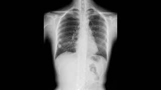
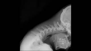
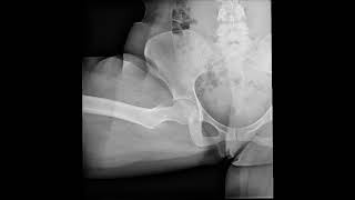
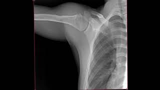
Dynamic Digital Radiography (DDR) is an advanced imaging technology that captures anatomy in motion. Unlike conventional X-rays that show only a single image, DDR provides dynamic visual information through a series of low-dose images displayed as a motion sequence. By visualizing movement, flow, and functional changes, DDR supports clinical assessment across a wide range of examinations while maintaining the simplicity and accessibility of standard radiography.
Seamless Integration with Konica Minolta Systems.
Native Compatibility: Designed for seamless integration with Konica Minolta systems, ensuring a hassle-free setup.
Uninterrupted Workflow: Access dynamic imaging data directly within your trusted Konica Minolta environment.
Smart Data Synergy: Smooth synchronization that bridges the gap between conventional radiography and advanced motion sequences.
Enabling DDR in mobile X-ray exam environments.*1
Frame Rate : 15fps.
Exposure Time : 20 seconds at longest.
*1 AeroDR 3 1417HD2 or AeroDR 1717HD2 is necessary for DDR
Clinical Application of DDR Technology
Musculoskeletal & Orthopedic Applications
Upper Extremities
Lower Extremities
Cervical & Lumbar Spine Stability
C-spine & L-spine
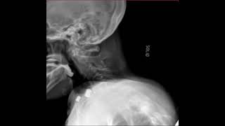
Swallow, Speech Pathology, and Post-Bariatric Sleeve Studies
Thoracic DDR & Dynamic Chest Radiography
Increasing Clinical Value with DDR
Operational Benefits
- Efficient Workflow: The system acquires up to 20 seconds of physiological movement. These exams can be performed by standard radiology staff without a physician’s presence, maximizing departmental throughput.
- Low-Dose Patient Safety: Provide dynamic imaging at a significantly lower radiation dose compared to fluoroscopy—making it suitable for pediatric patients and follow-up exams.
- Flexible Imaging: Enable imaging in various positions, including standing, seated, and supine, allowing clinicians to replicate real-life symptoms.
Expanding Clinical Applications
- Musculoskeletal & Functional Assessment: Visualizes joint motion and spinal stability beyond static imaging, helping identify causes of pain, instability, and abnormal movement.
- Swallowing Studies: Enables dynamic evaluation of swallowing function at low dose, supporting functional assessment.
- Contrast Studies: Captures motion during contrast-enhanced exams, supporting functional diagnosis across a range of clinical applications.
Clinical Value
- Treatment Follow-up: Enables evaluation of postoperative and follow-up motion before proceeding to advanced modalities such as CT or MRI.
- Improved Communication: Motion visualization enhances patient understanding and supports more effective communication between clinicians and patients.
The Experts Speak
DDR Uses In Contrast Examination
DDR Uses in Orthopedics
More Product Information
Learn more about the product in the brochure.